Injury to the Ligaments & Meniscus During Sports

The ligaments and the meniscus are structures within and around the knee joint. They and the cartilage of the end of each bone are structures involved in supporting and cushioning the joint. These structures themselves have a limited blood supply and do not necessarily heal well as a result. Bones and muscles, by comparison, tend to have a rich blood supply and may replenish or scar to nearly as good as new with little more than rest and especially with rehabilitation.
Meniscal Tears
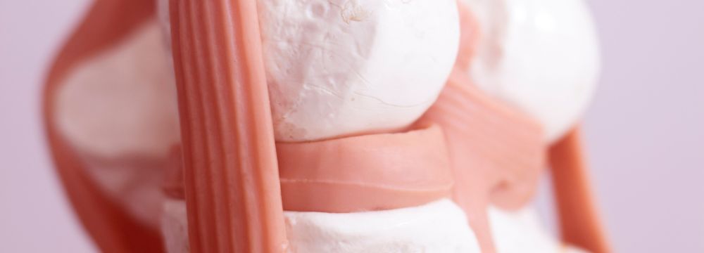
The meniscus is an extension of ligament-type tissue around the perimeter of the knee joint between the distal femur and proximal tibia. It appears like a gasket at the inner and outer areas where the bones in the knee meet.
Sometimes rest and anti-inflammatory regimens (by medications such as ibuprofen and ice/heat regimens) alone may improve meniscal injuries. Still, trimming may occasionally be required to shave away frayed edges, which continue impinging between joint ends. Depending on the location and severity of a tear (often appearing as or described as a ‘bucket handle’), the injured meniscus may benefit from a tacking repair. Alternately, or with failure of repair (often due to the limited blood supply and strain of rehab and recurrence), removal of significant portions of the meniscus may be the only way. This, unfortunately, can lead to advanced arthritis (bone-on-bone stress and wear) of the knee and eventually a knee replacement.
What About the Ligaments?
The lateral (outer) and medial (inner) collateral ligaments offer supportive stability, mostly side-to-side for the knee from outside the joint. At the same time, the anterior and posterior ligaments provide stability mostly front-to-back but also side-to-side from within the joint. The inner ligaments are named ‘cruciate’ as they cross one another at the interior compartment of the knee. The ACL (anterior cruciate ligament) extends from the distal femur’s lateral posterior (outside and behind) and crosses from behind to the front of the knee. It crosses in front of the PCL (posterior cruciate ligament). Twisting, pivoting, and abrupt changes of direction or momentum stress all these structures, which are elastic to a point and then tear or rupture completely. Understanding the exact motion (mechanism of injury) can help focus exams and diagnostics on which and/or how many of these structures may have been injured.
These cruciate and collateral ligaments may be partially torn within their length or completely disrupted from their bone attachments. With more significant stresses often seen in sports collisions or collapses, the failure of these ligaments can also disrupt and tear blood supply structures, which can have catastrophic results. Even hours after such an injury, it is essential to monitor pulse flow below the knee, at the ankle, and at the foot to be sure these blood vessels have not been torn or compressed by swelling within the knee in a condition known as Compartment Syndrome. A decrease in pulse below the knee, especially in distal numbness, tingling, or whitened skin tone for any reason, is considered a surgical emergency.
The severity of the injury will, of course, determine the appropriate course of action moving forward. Importantly, visiting an experienced orthopedic surgeon is crucial to understanding if other injuries if it’s sustained beyond those that are most obvious. The proper treatment of ligament injuries in and around the knee is critical to long-term function, from rest and home therapies to physical therapy and ultimately reconstructive surgery.
The team here at Premier Ortho is dedicated to seeing patients as soon as possible after an injury to offer the most effective treatment possible. Don’t hesitate to contact our office for more information and an evaluation.


 ES
ES